SVT vs. VT: Understanding the Difference Between Supraventricular and Ventricular Tachycardia


The Short Answer: SVT starts at or above the AV node and usually produces a fast, narrow-complex rhythm, while VT starts in the ventricles and produces a wide-complex rhythm that can be life-threatening. Telling them apart quickly guides treatment, because a drug that helps SVT can be dangerous in VT.
Supraventricular tachycardia and ventricular tachycardia can look alike at a glance and share symptoms like palpitations, chest pain, and dizziness. But they come from different parts of the heart, carry different risks, and call for different treatment.
What Is Supraventricular Tachycardia (SVT)?
SVT is a fast heart rate that begins at or above the AV node, in the heart's upper chambers or the AV junction. Because the electrical signals still travel through the normal conduction system, the QRS complexes are usually narrow.
Common forms of SVT include:
- Paroxysmal supraventricular tachycardia (PSVT): sudden episodes that start and stop on their own, often from a reentry loop near the AV node
- AVRT: uses an extra accessory pathway between the atria and ventricles
- Atrial tachycardia: a fast focus in the atria firing above the sinus node
- Atrial flutter and atrial fibrillation: organized flutter waves or a disorganized atrial rhythm
Sinus tachycardia is different. It is the sinus node speeding up a normal rhythm in response to exercise, fever, or stress, not an abnormal circuit. Most forms of SVT are not immediately dangerous, though frequent episodes can affect quality of life and, over time, strain the heart.
What Is Ventricular Tachycardia (VT)?
VT is a fast rhythm of three or more beats above 100 beats per minute that starts below the AV node, in the ventricles. Because the signal skips the normal conduction path, the QRS width is wide.
VT comes in two main patterns:
- Monomorphic VT: a single ventricular focus produces QRS complexes of one uniform shape
- Polymorphic VT: the QRS shape varies beat to beat, as seen in torsades de pointes
VT is the more dangerous rhythm. It often appears in patients with heart disease, such as a prior myocardial infarction or heart failure, and it can deteriorate into ventricular fibrillation and cardiac arrest. That risk is why VT is treated with urgency, even when a patient looks stable at first.
SVT vs VT: The Core Differences
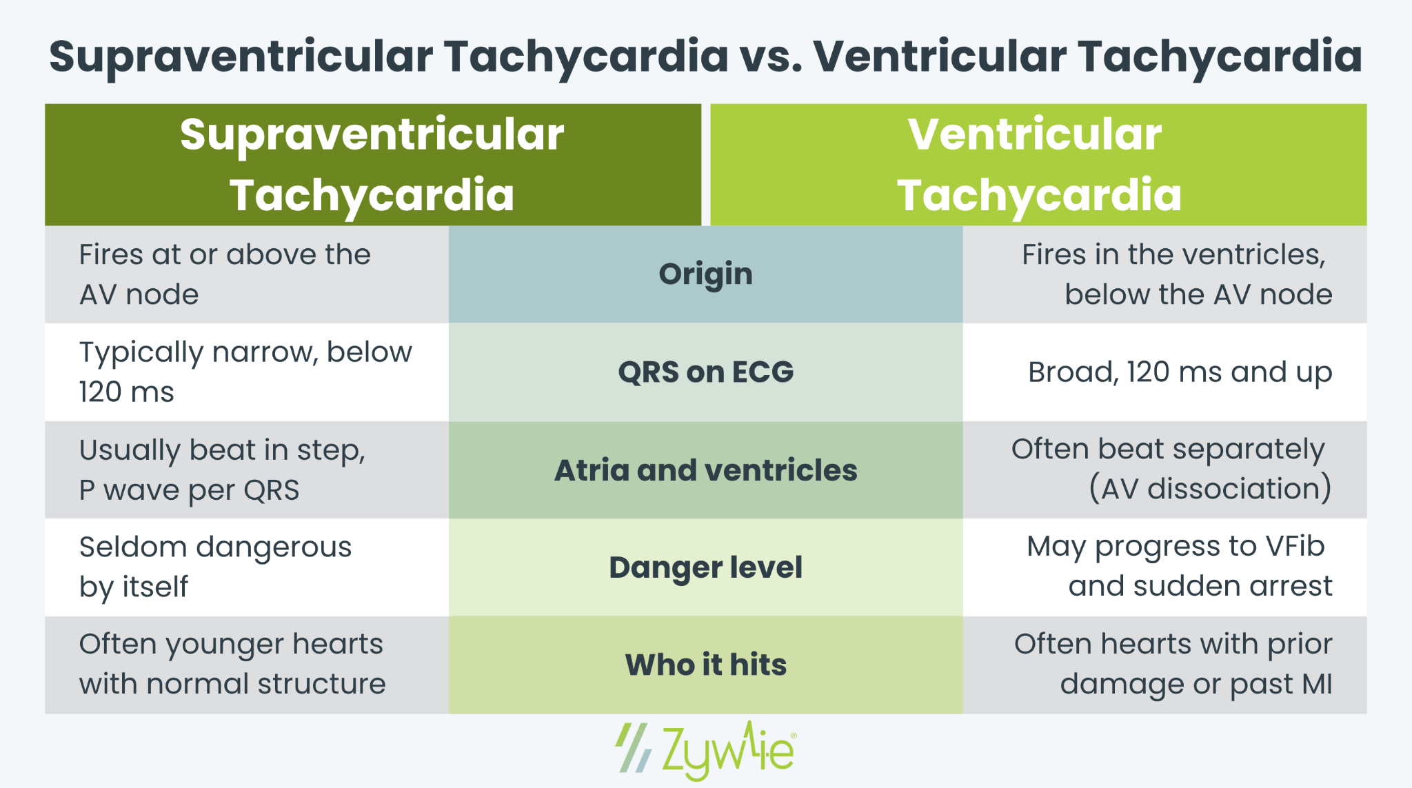
The two rhythms separate along a handful of features. Here is how they compare:
- Where it starts: SVT begins at or above the AV node. VT begins below it, in the ventricles.
- QRS width: SVT is usually narrow, under 120 ms. VT is wide, 120 ms or more.
- Rhythm: SVT is often a regular rhythm. VT is regular or slightly irregular.
- P waves: in SVT, a P wave usually ties to each QRS. In VT, the atria and ventricles often beat on their own, a pattern called AV dissociation.
- Risk level: SVT is rarely life-threatening on its own. VT can decay into ventricular fibrillation and cardiac arrest.
- Typical patient: SVT often shows up in younger, structurally normal hearts. VT is more common in patients with structural heart disease or a prior myocardial infarction.
The Wide Complex Problem
Here is where the two overlap and where mistakes happen. SVT usually shows a narrow complex, but it can produce a wide QRS when conduction is abnormal, a situation called aberrant conduction, often from a bundle branch block or an accessory pathway. When that happens, SVT looks like VT on the monitor. Both fall under the label of wide complex tachycardia.
The odds still favor VT. Studies show that about 80% of wide complex tachycardias are VT, and the figure climbs above 90% in patients with a prior myocardial infarction. ACLS guidelines reflect this: treat an undifferentiated wide complex tachycardia as VT until proven otherwise.
The reason is safety. Giving an AV nodal blocker such as a calcium channel blocker, for example, verapamil, to a patient who actually has VT can cause a sharp drop in blood pressure and trigger cardiac arrest. Treating SVT as VT is usually the safer error, which is why a cautious read is the default.
How Clinicians Tell Them Apart
No single clue is perfect, so clinicians weigh several together.
Clinical History
A history of heart disease, prior myocardial infarction, or heart failure points strongly toward VT. Age and symptoms help less than many expect, since a stable, comfortable patient can still be in VT.
ECG Features That Favor VT
- AV dissociation: the atria and ventricles beat independently, the most specific sign of VT
- Capture or fusion beats: occasional normal-looking beats breaking through the wide complex
- Concordance: all precordial leads point in the same direction
- A very wide QRS with certain LBBB morphology or right bundle branch block shapes
The Brugada Criteria
The Brugada criteria are a four-step ECG method for separating VT from SVT with aberrant conduction. Each step checks a feature, such as the absence of an RS complex, a long R-to-S interval, or AV dissociation, and if any step points to VT, the analysis stops there. The original method reported high accuracy, though real-world numbers run lower, so it guides judgment rather than replacing it.
How SVT and VT Are Treated
Treatment depends on the rhythm and on whether the patient is stable.
SVT:
- Start with a vagal maneuver, such as bearing down
- Adenosine to interrupt the reentry loop
- Beta blockers or a calcium channel blocker to slow the ventricular rate
- Catheter ablation for recurrent PSVT or an accessory pathway
VT:
- An unstable patient gets synchronized electrical cardioversion right away
- A stable monomorphic VT patient may receive IV antiarrhythmic drugs
- Catheter ablation for recurrent VT and an implantable defibrillator for patients at ongoing risk of cardiac arrest
If any patient is unstable, with low blood pressure or severe chest pain, the rhythm label matters less in that moment. Electrical cardioversion comes first, in line with ACLS guidelines. Once the rhythm settles, the goal shifts to finding the cause and preventing the next episode.
Why Capturing the Rhythm Matters
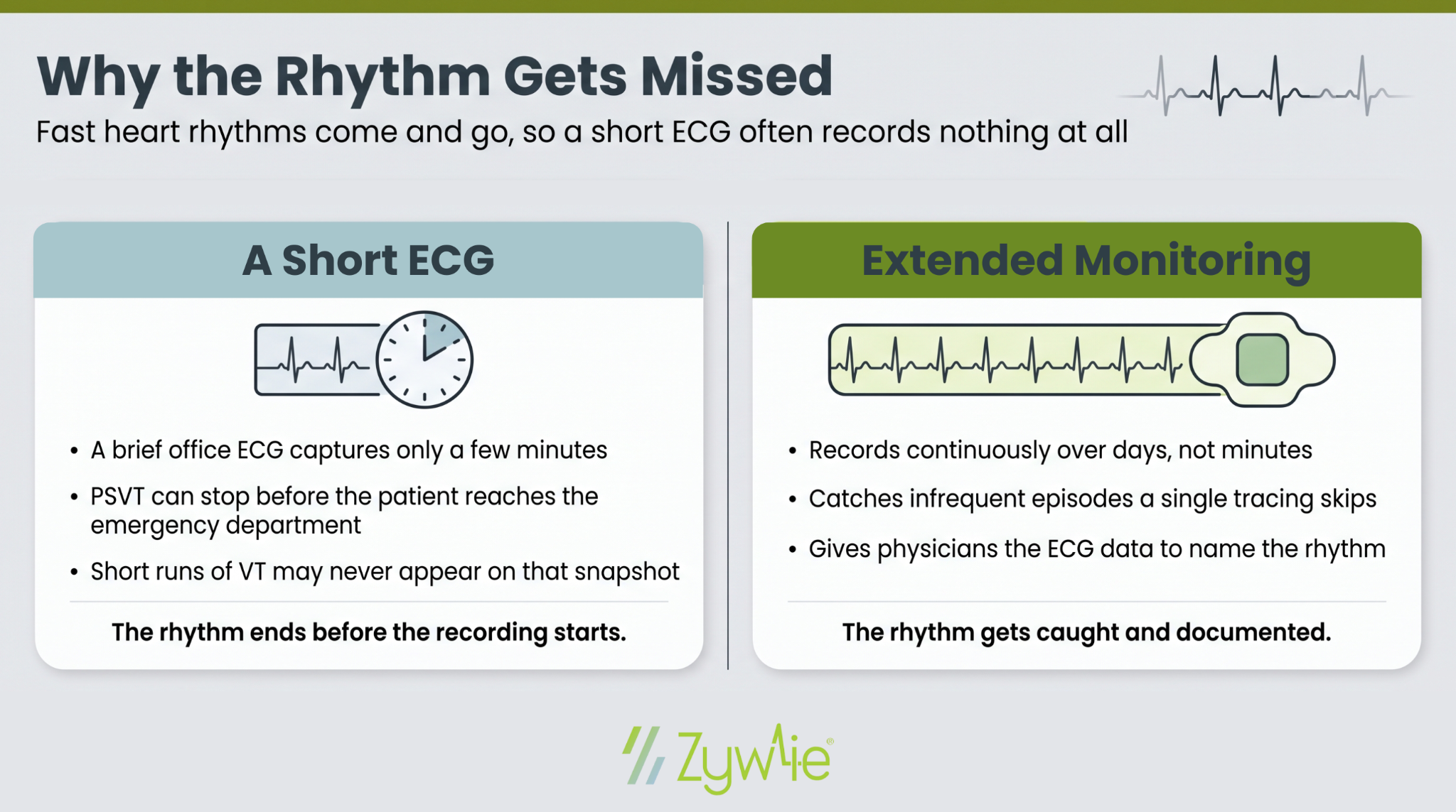
Many of these rhythms come and go. Paroxysmal supraventricular tachycardia can stop before a patient reaches the emergency department, and short runs of VT may never appear on a brief office ECG. If the rhythm is not recorded, the diagnosis stays a guess, and treatment waits with it.
Extended cardiac monitoring closes that gap. Recording the heart over days captures the abnormal heart rhythms a single tracing misses and gives physicians the ECG data to name the rhythm and return the patient to normal sinus rhythm with the right therapy. Longer monitoring windows also raise the chance of catching infrequent episodes that a 24-hour test would skip.
Getting to the Right Diagnosis Faster
SVT and VT can mimic each other, but the difference decides both the treatment and the level of risk. A wide complex rhythm deserves caution, a careful look at the ECG, and often a longer recording to catch what a snapshot cannot.
That is where remote monitoring helps. Zywie Healthcare's ZywieNano™ captures up to 30 days of continuous ECG data and transmits it to a clinician -reviewed monitoring center in near real-time, so an arrhythmia reaches the care team without waiting for a device to come back and be analyzed. Physicians get clinically accurate data and timely alerts that help separate SVT from VT and act sooner.
Contact Zywie Healthcare to learn how continuous ECG monitoring can support faster, more confident rhythm diagnoses.
Join Us in Revolutionizing Cardiology – Let's Transform Heart Health Together