PVC Burden: What It Means and How Heart Monitoring Measures It

The Short Answer: PVC burden is the percentage of your total heartbeats that are premature ventricular contractions over a set recording period. A burden under 1% is common and usually harmless, while a burden above 10% raises the risk of PVC-induced cardiomyopathy and generally calls for closer follow-up.
Nearly everyone has a few extra beats. What separates a harmless finding from one that needs treatment is how often the beats occur and what they are doing to the left ventricle over time. That is what PVC burden measures.
What Is a PVC?
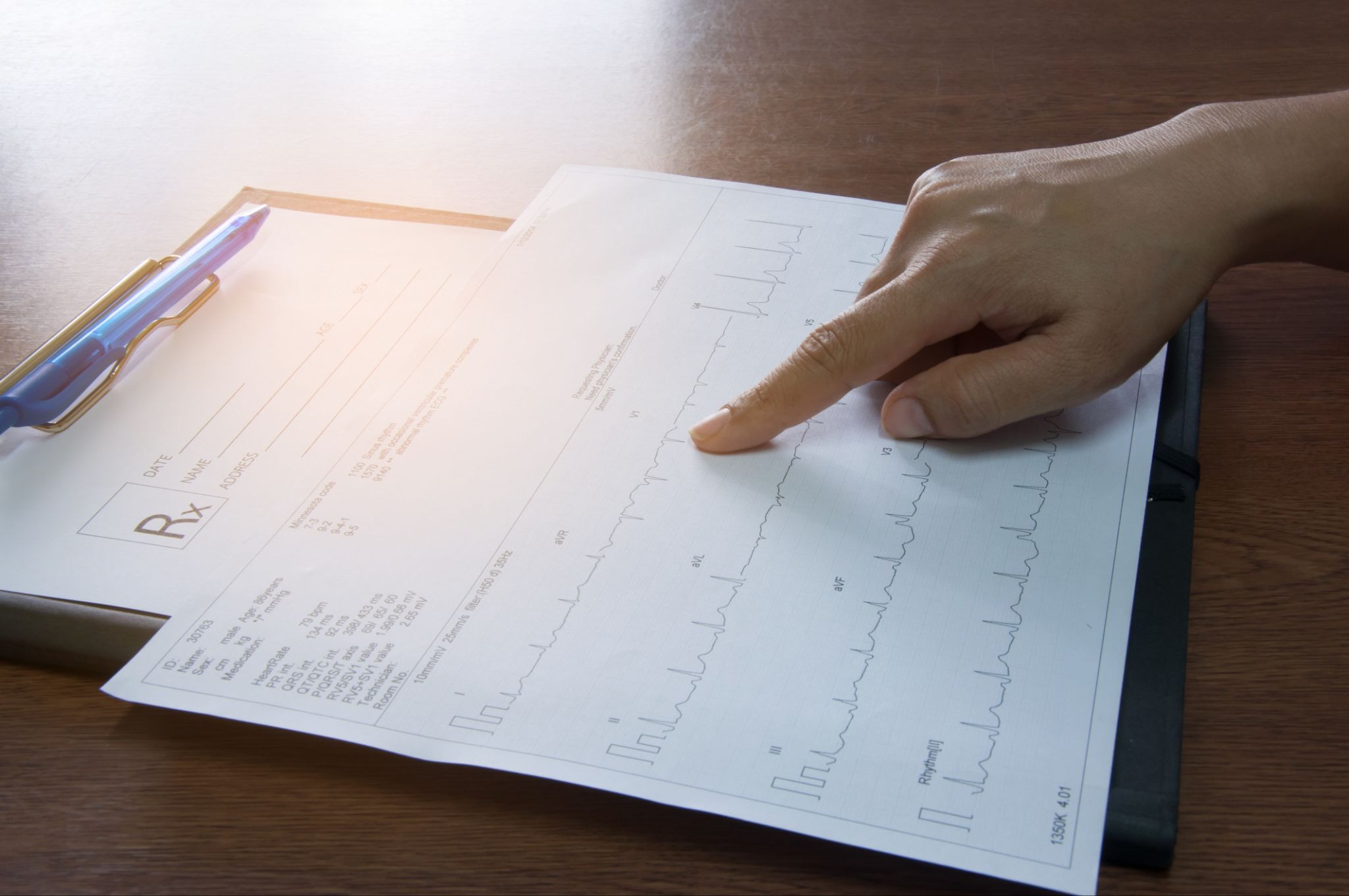
A premature ventricular contraction is an early heartbeat that starts in the ventricles instead of the sinus node. Because the signal skips the normal conduction path, the beat arrives early and the QRS complex looks wide and oddly shaped on a 12-lead ECG.
Most people feel nothing. Those who do describe a skipped beat, a thud, or a flutter in the chest, which comes from the stronger beat that follows the pause rather than the PVC itself.
PVCs are also called premature ventricular complexes, ventricular extrasystoles, or simply extra beats. For a closer look at how they differ from their atrial counterpart, see our breakdown of PACs versus PVCs.
How PVC Burden Is Calculated
PVC burden is a straightforward ratio:
PVC burden = (number of PVCs ÷ total heartbeats) × 100
If a patient records 100,000 beats over 24 hours and 12,000 of them are PVCs, the burden is 12%.
The math is simple. The complication is the denominator. A 24-hour Holter monitor captures roughly 100,000 beats. A 14-day patch captures more than a million. The longer the window, the more the number reflects a true daily average rather than one unusual day.
What the Numbers Mean
There is no single cutoff that applies to every patient, but clinical practice generally groups burden into these ranges:
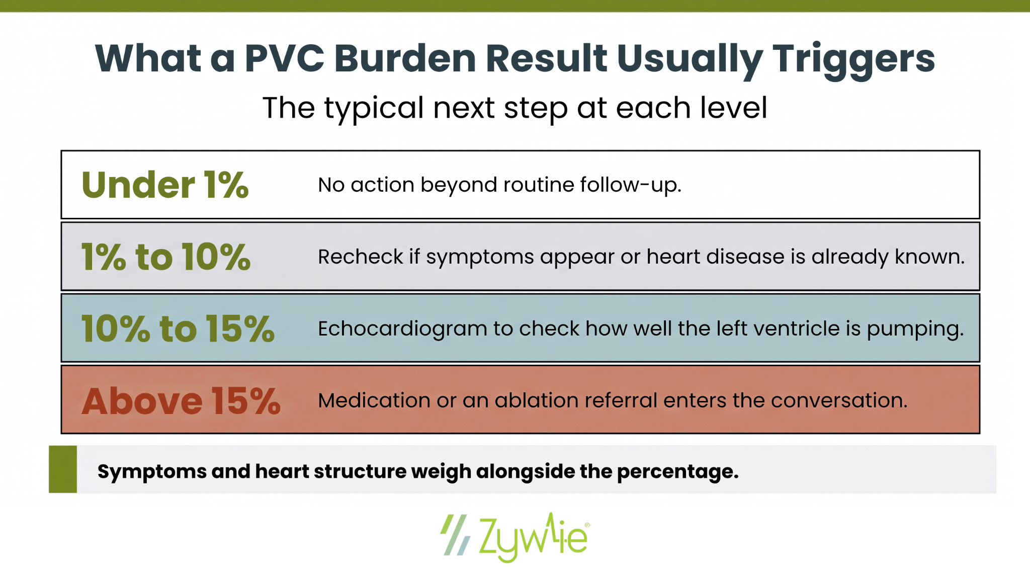
- Under 1%: Occasional PVCs. Common in healthy hearts and usually left alone.
- 1% to 10%: A moderate burden. Often monitored, especially if symptoms are present or the patient has structural heart disease.
- 10% to 15%: The zone where studies begin to show a link to reduced left ventricular ejection fraction. Follow-up imaging is typical.
- Above 15% to 20%: A high PVC burden with a meaningful risk of PVC-induced cardiomyopathy. Treatment is usually discussed.
Those lines trace back to a study of 174 patients referred for PVC ablation. A burden above 24% best separated patients with impaired left ventricular function from those with preserved function, and the lowest burden that produced a reversible cardiomyopathy was 10%.
Burden is one input, not the whole picture. A patient with a 6% burden and coronary artery disease may warrant more attention than a patient with a 14% burden and an otherwise normal heart.
Why High PVC Burden Matters
PVC-Induced Cardiomyopathy
When a large share of beats originate in the ventricle, the heart pumps less efficiently. Over months to years, that inefficiency can weaken the left ventricle and lower ventricular ejection fraction, a condition known as PVC-induced cardiomyopathy or arrhythmia-induced cardiomyopathy.
The important detail: it is often reversible. When the PVC burden drops through medical therapy or ablation, LV function frequently recovers.
Signals of Higher Risk
Not all PVCs carry the same weight. Features that raise concern include:
- A wide PVC QRS duration, which suggests the beat originates farther from the normal conduction system
- PVCs that come from multiple sites rather than one focus
- Runs of consecutive PVCs, which can progress toward ventricular tachycardia
- An underlying structural heart problem, prior heart failure, or known coronary artery disease
- Symptoms such as chest pain, lightheadedness, or fainting
In a structurally normal heart, most PVCs are idiopathic and carry a low risk of ventricular fibrillation. In a damaged heart, the same finding is weighed differently.
Where PVCs Come From
The right ventricular outflow tract is the most common origin for idiopathic PVCs. Beats from the outflow tracts often produce a recognizable pattern on ECG, which helps electrophysiologists plan a PVC ablation before the procedure begins.
Why Recording Length Changes the Answer
PVC burden fluctuates. It shifts with sleep, exercise, caffeine, stress, and time of day. A single 24-hour snapshot can land well above or well below a patient's true average.
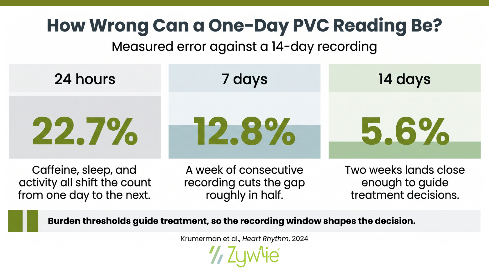
A large national cohort study quantified the gap. Compared with 14-day continuous monitoring, a single 24-hour period produced a 22.7% measured error in PVC burden. Extending to 7 consecutive days cut that to 12.8%, and 14 days brought it down to 5.6%.
That variability creates two problems:
- Underestimation: A quiet recording day can suggest a low burden and delay treatment for someone who actually sits above the threshold.
- Overestimation: An unusually busy day can push a patient into a treatment conversation they may not need.
Extended monitoring narrows the error. Recording over 7, 14, or 30 days averages across good days and bad ones, and it also reveals patterns a short study misses, such as burden that spikes overnight or only during activity. That distinction matters because a burden trending upward over weeks calls for a different plan than one holding steady.
How PVC Burden Is Managed
Watchful Waiting
For asymptomatic PVCs with a low burden and a normal echocardiogram, no treatment is needed. Periodic monitoring confirms the burden is not climbing.
Medical Therapy
Beta blockers are the usual first-line treatment, particularly when symptoms are the main complaint. A calcium channel blocker is an alternative for patients who cannot tolerate beta blockade. Both reduce symptoms reliably but often produce only a modest drop in burden.
Removing triggers matters too. Certain medications, including decongestants and antihistamines, along with alcohol, stimulants, and untreated sleep apnea, can all raise PVC frequency.
Catheter Ablation
For a high PVC burden, especially with declining ejection fraction or symptoms that persist despite medication, catheter ablation targets the specific focus producing the extra beats. Radiofrequency ablation of a single outflow tract focus has strong success rates and a low rate of adverse events in experienced hands.
Ablation is often the better choice when PVC-induced cardiomyopathy is suspected, since reducing the burden is what allows LV function to recover.
Additional Testing
Cardiac magnetic resonance imaging is sometimes used to look for scar or infiltrative disease that an echocardiogram cannot see. It helps separate PVCs that caused a cardiomyopathy from PVCs that are a symptom of an underlying heart condition.
Getting an Accurate Burden Number
PVC burden is a percentage, and percentages are only as good as the sample behind them. A one-day recording answers what happened on one day. A multi-week recording answers what is happening to the patient.
That difference decides real things: whether a patient gets an echocardiogram, whether a beta blocker gets started, and whether an ablation conversation happens now or in six months.
Zywie Healthcare's ZywieNano™ patch records continuously for up to 30 days with beat-to-beat analysis, transmitting data to a physician-reviewed monitoring center in near real-time. Clinicians get burden trends across the full window rather than a single-day estimate, with timely alerts when findings warrant attention.
Explore the ZywieNano™ device suite or contact Zywie Healthcare to see how extended monitoring supports faster, more confident PVC decisions.
Join Us in Revolutionizing Cardiology – Let's Transform Heart Health Together