ECG vs. EKG vs. Echocardiogram: What's the Difference?


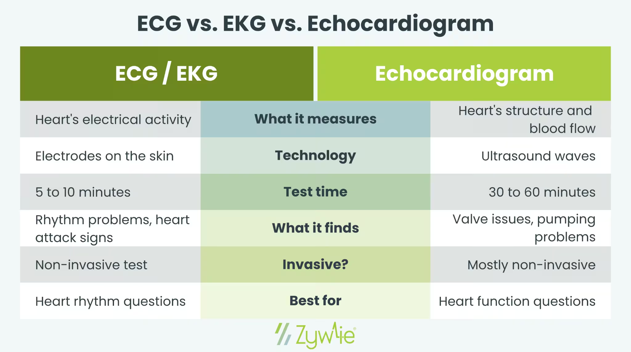
The Short Answer: An ECG and EKG are the same test; both record the heart's electrical activity to detect rhythm problems. An echocardiogram uses sound waves to create images of the heart's structure and how blood flows through it.
If a doctor recommends one of these tests, it helps to know what makes each one different. ECG, EKG, and echocardiogram all check heart health, but they look at very different things. An ECG records electrical signals from the heart muscle. An echocardiogram shows what the heart looks like and how blood moves through it.
What Is an ECG?
An ECG, also called an electrocardiogram, records the heart's electrical activity through small electrodes placed on the chest, arms, and legs. It is one of the most common diagnostic tools used to check heart function. A standard ECG test takes about 5 to 10 minutes in a doctor's office.
How an ECG Works
- Small sticky pads called electrodes attach to the skin
- Each electrode picks up the electrical signal that triggers a heartbeat
- A machine records these electrical impulses as wavy lines on paper or a screen
- A healthcare provider reviews the ECG results to spot any abnormal heart rhythm
What an ECG Can Detect
According to the Mayo Clinic, an ECG can help identify:
- Atrial fibrillation and atrial flutter
- Slow or fast heart rate
- Signs of a past heart attack or myocardial infarction
- Ventricular hypertrophy (thickened heart muscle)
- Irregular heartbeat patterns
Is ECG the Same as EKG?
Yes. ECG and EKG refer to the same test. The difference comes from language. EKG comes from the German word "elektrokardiogramm," while ECG is the English version. American doctors often use an EKG to avoid confusion with an EEG, which records brain activity. Both terms describe a non-invasive test that measures the heart's electrical activity.
What Is an Echocardiogram?
An echocardiogram, often called an echo or echo test, uses ultrasound waves to create moving pictures of the heart. Instead of recording electrical activity, an echo test shows the heart's structure, valves, and the blood flow through each chamber.
How an Echo Works
A technician places an ultrasound probe on the chest. The probe sends sound waves that bounce off the heart muscle and return as images on a screen. The whole process usually takes 30 to 60 minutes.
Types of Echocardiograms
- Transthoracic echocardiogram: The most common type. The ultrasound probe moves across the outside of the chest.
- Transesophageal echocardiogram: A small probe passes down the throat for a closer view. Used when more detail is needed.
- Stress echocardiogram: Done with a stress test to see how the heart works during exercise.
- Doppler ultrasound: Measures the speed and direction of blood flow through the heart and vessels.
What an Echo Can Detect
- Heart valve problems
- Structural problems with the heart muscle
- Signs of heart failure
- Blood clots inside the heart
- Damage from a recent heart attack
- Fluid buildup around the heart
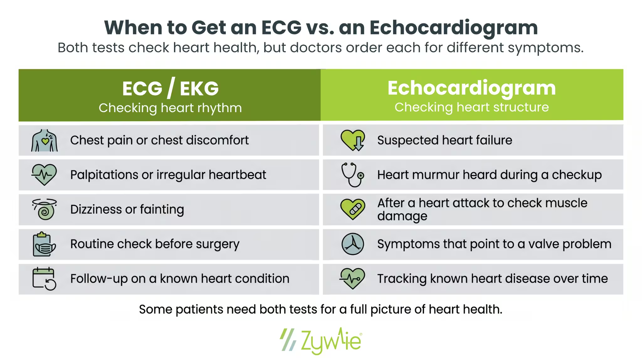
When Doctors Order Each Test
Doctors use ECGs and echocardiograms to look at different parts of heart health. An ECG is often used to check the heart’s rhythm and electrical activity, while an echocardiogram gives a closer look at the heart’s structure, valves, and pumping function.
Here are some common reasons a doctor mat order each test:
What About Longer Heart Monitoring?
A standard ECG only captures a few minutes of data. For patients with symptoms that come and go, doctors may order a longer monitoring period using:
- Holter monitor: Records continuously for 24 to 48 hours
- Event monitor: Patient activates it when they feel symptoms
- Mobile cardiac telemetry (MCT): Records and transmits ECG data for up to 30 days while patients carry on with normal activities
Extended monitoring catches abnormal heart rhythm patterns that a 10-minute test might miss. Research published in The New England Journal of Medicine found that continuous monitoring detected atrial fibrillation in 6.4 times more patients than standard care at six months.
For a deeper look at extended monitoring, see Zywie's guide on What Is an MCT Monitor.
Other Heart Tests You May Hear About
Doctors sometimes pair these tests with other diagnostic tools to reach an accurate diagnosis:
- Coronary angiogram: An imaging test that looks at the coronary arteries for blockages. A doctor injects dye through a thin tube to make the arteries show up clearly on an X-ray.
- Cardiac MRI: Uses magnets and radio waves to create detailed images of the heart muscle, valves, and major vessels. Often used when an echo does not give enough detail.
- Stress test: Measures heart performance during exercise on a treadmill or stationary bike. Doctors may pair this with an ECG or echo to see how the heart responds to physical activity.
Many patients with suspected heart disease end up needing more than one test to get a full picture. For example, an ECG might catch an irregular heartbeat, while an echo shows whether the heart is pumping well. Combining results gives the care team a clearer view before recommending treatment.
Preparing for Each Test
Before an ECG
- Wear loose, comfortable clothing
- Skip lotions or oils on the chest
- Tell the technician about any medications
Before an Echo
- No special prep for a standard transthoracic echocardiogram
- Skip food and drinks for several hours before a transesophageal echocardiogram
- Plan for a ride home if sedation is involved
Understanding Your Results
After an ECG or echo, a healthcare provider reviews the data and explains the findings.
What a Normal ECG Shows
A normal ECG shows a steady heart rhythm with regular spacing between beats. The waves on the readout follow a predictable pattern that reflects the electrical impulse moving through the upper and lower chambers. A heart rate between 60 and 100 beats per minute at rest is generally considered normal for adults.
What an Echo Looks At
For an echo, providers look at how well each chamber pumps, whether the valves open and close correctly, and how blood flows through the heart. Some patients may need follow-up testing or longer monitoring if the results point to a possible heart condition that did not show up clearly in a short window.
How Zywie Healthcare Supports Better Heart Monitoring
ECGs and echocardiograms each play a role in heart health, but neither captures what happens day to day. For patients with intermittent symptoms like palpitations or shortness of breath, long-term cardiac monitoring fills the gap between office visits.
Zywie Healthcare offers the ZywieNano™, a small wearable that records continuous ECG data for up to 30 days. Data is transmitted in near real-time, so providers receive timely alerts when an abnormal heart rhythm appears. That shortens the path to an accurate diagnosis for conditions like atrial fibrillation and atrial flutter.
For providers looking to add extended cardiac monitoring to their workflow, or patients curious about long-term options beyond a standard ECG, contact Zywie Healthcare to learn more.
Join Us in Revolutionizing Cardiology – Let's Transform Heart Health Together
