How AI ECG Interpretation Supports Heart Monitoring

The Short Answer: AI ECG interpretation uses trained algorithms to read heart tracings, flag an abnormal ECG, and help clinicians act faster. It works best as a support tool that speeds arrhythmia detection and triage, while a certified clinical monitoring technician confirms the specific diagnosis.
Reading an ECG takes skill, and even experienced clinicians can miss subtle patterns during a busy shift. Artificial intelligence now assists by scanning ECG data in seconds and pointing to findings that need a closer look. For cardiac monitoring, that speed matters.
What AI ECG Interpretation Actually Does
AI ECG interpretation applies computer models to the electrical signals recorded by an ECG. The idea is straightforward: read the tracing, compare it against patterns learned from large sets of ECG data, and return a result the care team can use.
Most systems handle three jobs:
- Separate an abnormal ECG from a normal one
- Point toward a specific diagnosis, such as atrial fibrillation or a bundle branch block
- Surface hidden ECG features that the human eye cannot see, like early signs of ventricular systolic dysfunction
The result is a faster first read. Instead of waiting in a queue for manual review, a tracing can be scored in seconds and routed to the right person.
How AI Reads a Heart Tracing
The technology behind most tools is deep learning, a branch of artificial intelligence that learns from examples rather than fixed rules. Developers feed a model thousands or millions of labeled tracings, and the model learns which patterns match which conditions.
A convolutional neural network, or CNN, is the most common design for ECG analysis. It scans a tracing the way it scans an image, learning which shapes and intervals line up with a given diagnosis. Other tools use artificial neural networks and deep neural networks trained the same way, on large volumes of ECG data.
What Can AI Detect on an ECG?
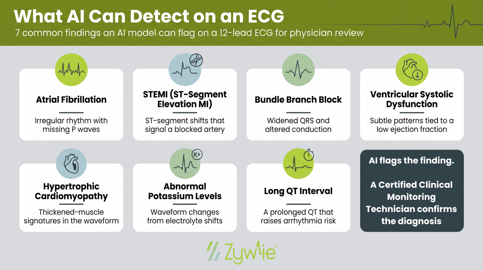
- Atrial fibrillation: a shaky, disorganized baseline with no steady P waves. AI is good at catching short episodes in long recordings, and early flags matter since AFib raises stroke risk.
- ST-segment elevation myocardial infarction (STEMI): an ST segment sitting higher than it should, a sign of a blocked artery. A quick flag moves a time-sensitive case up the queue.
- Bundle branch block: a QRS wider than normal, showing the signal is taking a delayed route through the lower chambers.
- Ventricular systolic dysfunction: faint patterns that line up with weak pumping, or low ejection fraction, often with no symptoms the patient notices.
- Hypertrophic cardiomyopathy: waveform marks that hint at a thickened heart wall, a condition tied to sudden cardiac events in younger people.
- Abnormal potassium levels: electrolyte swings leave a mark, like tall, pointed T waves, that a model can surface fast.
- Long QT interval: a longer-than-normal reset between beats that can trigger dangerous rhythms, sometimes from certain medications.
How Accurate Is AI ECG Interpretation?
Accuracy depends on the task. Broad checks are strong. Naming an exact condition is harder.
- A Mayo Clinic convolutional neural network detected low ejection fraction, a sign of ventricular systolic dysfunction, with an AUC of 0.93 and about 85.7% accuracy when tested on more than 52,000 patients, according to a study in Nature Medicine. AUC is a score where 1.0 is perfect.
- Specialized deep neural networks built for one job, such as arrhythmia detection or ST-segment elevation, often report higher accuracy above 90%.
- A diagnostic accuracy study of GPT-4o published in JMIR AI found it recognized ECG images with 100% accuracy and sorted normal from abnormal ECGs at 83% accuracy. Its classification accuracy for a specific pathology fell to 41%, showing the gap between screening and diagnosis.
- One health-system analysis reported by GE HealthCare found AI-assisted reading identified a major ECG abnormality more often than the conventional algorithm, 92.2% versus 87.2%
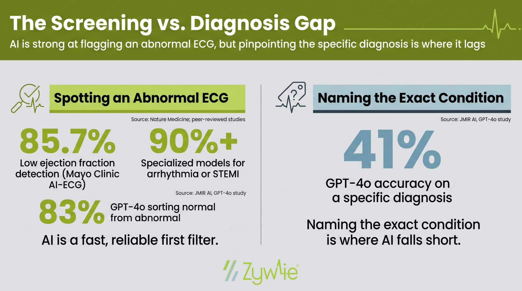
The pattern is clear. AI is a strong first filter for an abnormal ECG. Multiclass classification into a specific diagnosis still trails human experts, which is why the strongest results come from models trained for a single, narrow task.
Where AI Helps in Real Clinical Settings
AI ECG interpretation earns its place in a few practical spots.
Faster Triage in the Emergency Department
An AI model can flag an abnormal tracing within seconds, helping emergency physicians see high-risk patients first. Faster reads mean fewer delays in care and quicker treatment decisions when minutes count.
Early Detection of Silent Disease
Screening routine ECGs can surface heart failure risk or ventricular systolic dysfunction before symptoms appear. That kind of early detection gives clinicians a head start on treatment that may otherwise wait until a patient feels unwell.
A Second Read for Non-Specialists
Clinics without a cardiologist on site can use an AI algorithm as a backup read, giving healthcare professionals added confidence before referral. In outpatient and rural settings, that support can widen access to reliable ECG analysis.
Continuous Remote Cardiac Monitoring
AI reviews long stretches of ECG data and surfaces events for physician review. This is where AI ECG analysis and remote monitoring meet, and where much of the clinical application is heading. A model can watch for an arrhythmia around the clock and pass only the relevant findings to a human.
The Limits Every Care Team Should Know
AI adds speed, but it is not a finished answer. Medical professionals should weigh a few caveats before relying on any AI ECG output.
- False positives: Some AI algorithms over-call abnormal findings. A scoping review of outpatient AI ECG use noted a tendency for certain algorithms to overdiagnose, which can trigger extra echocardiograms and follow-up visits.
- A specific diagnosis is still hard: As the GPT-4o data shows, naming the exact condition lags well behind broad normal-versus-abnormal sorting.
- Support, not replacement: Most tools work as a support layer. A physician still owns the final diagnosis and treatment decisions, and any AI used at the point of care should meet medical device standards for clinical use.
- Data quality and bias: ECG features can differ across populations. A model trained on narrow ECG data may not carry the same high accuracy everywhere, which limits how far one AI model generalizes.
The takeaway for clinical practice: treat AI as a fast, reliable assistant that widens coverage, with a certified clinical monitoring technician or physician review anchoring every decision.
Turning AI-Ready ECG Data Into Action
AI ECG interpretation speeds the first read, flags abnormal ECGs, and helps care teams reach a diagnosis sooner. It performs best paired with clinical review, where the algorithm handles scale, and the clinician confirms the specific diagnosis.
That pairing is where remote monitoring fits. Zywie Healthcare's ZywieNano™ captures up to 30 days of continuous ECG data and transmits it to a certified cardiac -monitoring center in near real-time, so arrhythmias and other findings reach your team without waiting for a device to be returned. You get clinically accurate data and timely alerts, built for modern cardiac care.
Want to see how AI-powered insights and Certified Cardiac Monitoring Technician-reviewed monitoring can support your practice? Contact the Zywie Healthcare team to get started.
Join Us in Revolutionizing Cardiology – Let's Transform Heart Health Together