Comparing AFib and Atrial Flutter: A Clinical Guide

Atrial Fibrillation vs. Flutter:
Causes, Risks, and How Cardiac Monitoring Helps
At a Glance: Atrial fibrillation and atrial flutter are both abnormal heart rhythms that originate in the upper chambers of the heart, but they differ in how their electrical signals behave, how they appear on an ECG, and how they are treated. Both carry serious risks, including stroke and heart failure, and accurate diagnosis through extended remote cardiac monitoring is the first step toward effective management.
Atrial fibrillation and atrial flutter are two of the most frequently confused atrial arrhythmias in clinical practice. Both disrupt the normal electrical activity of the atria and can produce similar symptoms. But the similarities end there. Their causes, patterns, and treatment paths are distinct, and treating one as if it were the other leads to delays in care. This post breaks down the two conditions, their shared and unique risk factors, treatment approaches, and why getting the diagnosis right from the start matters.
What Is Atrial Fibrillation?
How AFib Works in the Heart
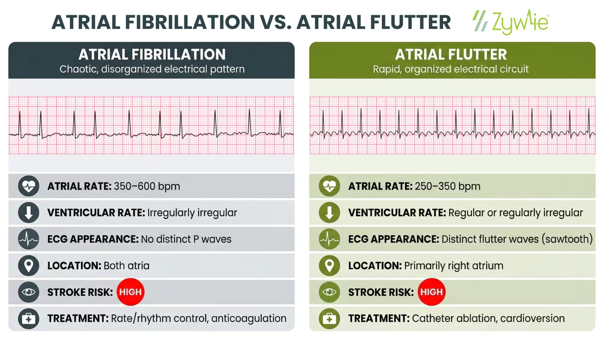
In atrial fibrillation, chaotic electrical signals fire rapidly throughout both atria with no organized pattern. Instead of contracting in a coordinated sequence, the atria quiver. This produces an irregular, often fast heart rate that typically runs between 100 and 175 beats per minute.
AFib is grouped into three types based on how long it lasts:
- Paroxysmal: Starts and stops on its own, often lasting less than 7 days
- Persistent: Lasts more than 7 days and requires treatment to stop
- Permanent atrial fibrillation: An ongoing rhythm that is no longer managed with rhythm restoration
Why AFib Is Dangerous
When the atria does not contract fully, blood can pool in the left atrial appendage and form a blood clot. If that clot travels to the brain, it causes an ischemic stroke. Long-term AFib is also associated with reduced ejection fraction, myocardial infarction, and progression toward heart failure.
The prevalence of atrial fibrillation has risen significantly over the past two decades, driven largely by aging populations and rising rates of cardiovascular disease, with estimates now running three times higher than projections made 20 years ago.
What Is Atrial Flutter?
How Atrial Flutter Works
In atrial flutter, a single electrical impulse loops repeatedly through a fast, stable circuit, most often within the right atrium. This creates a rapid but organized atrial rate of roughly 250 to 350 beats per minute.
The AV node acts as a gatekeeper, blocking most of those impulses before they can reach the ventricles. This keeps the ventricular rate at a more manageable level, typically 75 to 150 beats per minute, and often regular.
On an ECG, this pattern produces the characteristic flutter waves, a repeating sawtooth appearance that distinguishes it from other atrial arrhythmias.
There are two main forms:
- Typical atrial flutter: Uses a circuit around the tricuspid valve in the right atrium; the most common and most treatable form
- Atypical atrial flutter: Involves a different circuit, sometimes in the left atrium; more complex to diagnose and manage
Why Atrial Flutter Is Also a Concern
Despite its more organized pattern, atrial flutter carries many of the same risks as AFib. Blood clot formation, ischemic stroke risk, and heart failure can all occur. Patients with atrial flutter also frequently develop atrial fibrillation over time, which further raises long-term stroke risk.
Atrial Fibrillation vs. Atrial Flutter: Side-by-Side Comparison
Shared Risk Factors for Both Conditions
AFib and atrial flutter share a large overlap in underlying causes:
- Hypertension: Long-term high blood pressure causes atrial enlargement and electrical remodeling
- Heart failure: Elevated pressure and structural changes in the atria increase arrhythmia risk
- Diabetes: Associated with oxidative stress and autonomic changes that contribute to atrial arrhythmias
- Alcohol: Even moderate intake has been linked to increased risk of both AFib and flutter
- Age: Risk rises sharply after 60, and the age-standardized rate of both conditions increases with each decade
How Each Condition Is Treated
Treating Atrial Fibrillation
AFib treatment focuses on three areas: controlling heart rate, restoring sinus rhythm, and reducing stroke risk.
- Rate control: Beta blockers and calcium channel blockers slow the ventricular rate without converting the rhythm
- Rhythm control: Antiarrhythmic drugs or electrical cardioversion can restore normal sinus rhythm
- Anticoagulation: Reduces the chance of blood clot formation in the atrial appendage
- Catheter ablation: Targets the abnormal electrical pathways driving AFib; evidence from clinical trial data supports its use when antiarrhythmic drugs have not worked
Treating Atrial Flutter
- Electrical cardioversion: Often effective at restoring a normal heart rate in flutter
- Radiofrequency ablation: An ablation procedure that uses heat to interrupt the flutter circuit in the right atrium; studies show high long-term success rates for typical atrial flutter ablation
- Rate control and anticoagulation: Used when rhythm control is not the immediate priority, following a similar approach to AFib management
One significant difference between the two: typical atrial flutter responds to radiofrequency ablation at very high rates. AFib ablation is more technically complex, has a longer refractory period for healing, and often requires multiple procedures.
Why Accurate Diagnosis Is the Starting Point
AFib and atrial flutter can produce identical symptoms: palpitations, shortness of breath, fatigue, and chest discomfort. But treating one condition as the other delays effective medical care.
The distinction matters for several reasons:
- An ablation procedure designed for typical atrial flutter targets a specific circuit in the right atrium, while AFib ablation requires a different approach entirely
- Atrial tachycardia and atypical atrial flutter require different mapping strategies than typical flutter
- Decisions about antiarrhythmic drugs, anticoagulation, and electrical cardioversion timing all depend on identifying the correct arrhythmia first
A standard ECG captures only a brief window of cardiac activity. Paroxysmal AFib and intermittent flutter may not appear during that window at all. Even a 24 to 48-hour Holter monitor misses arrhythmias that occur less frequently.
Extended monitoring, from 3 to 30 days, significantly increases the likelihood of capturing a symptomatic episode. A systematic review of extended cardiac monitoring data consistently found that longer monitoring windows improve diagnostic yield across a range of atrial arrhythmias, leading to faster treatment decisions and better patient outcomes.
How Zywie Healthcare Supports Faster Diagnosis
Accurate, timely diagnosis is what makes every downstream treatment decision possible. When clinicians can pinpoint exactly what arrhythmia a patient has, they can move forward with confidence: selecting the right procedure, adjusting antiarrhythmic drugs, or calibrating anticoagulation based on real data rather than assumptions.
Zywie Healthcare provides remote cardiac monitoring built around that diagnostic need.
The ZywieNano patch delivers up to 30 days of continuous ECG data, transmitted in near real-time, allowing clinicians to receive timely alerts when a cardiac event is identified, without waiting for the device to be returned.
For practices managing patients with suspected atrial fibrillation, atrial flutter, or other atrial arrhythmias, faster and more accurate data means faster treatment and fewer delays in care. Visit the For Providers page to explore Zywie's full device suite, or contact the team to learn how Zywie fits into your monitoring workflow.
Join Us in Revolutionizing Cardiology – Let's Transform Heart Health Together
